Sunday, August 10, 2014

Tips and Pearls for Chest X Ray part II

- Air fluid level
  • Air fluid level in esophagus = esophageal stasis
  • Intrapulmonary cavity with air fluid level - upper walls must be visible
  • Hydropneumothorax = indistinct upper walls with different 'shape' of air fluid levels in PA and lateral view ( lower level can be outlined by the curved minor fissure )
- Thymoma can regularly metastasize to pleural layers

- Any opacity in diaphragm may arise below

- Lymph node with hypodense center - think about TB

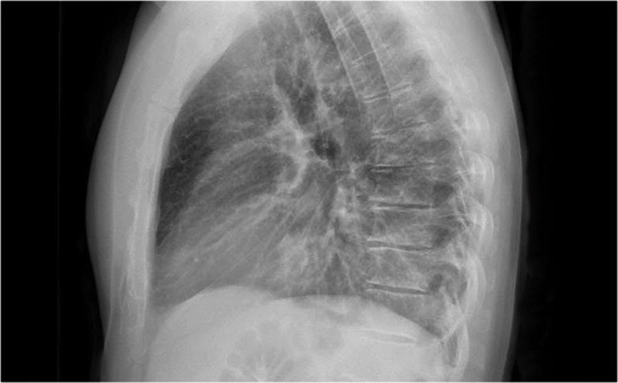
- Ivory vertebrae ddx - adult - malignancy, lymphoma, Paget's disease of bone; kids - lymphoma, neuroblastoma, osteoblastoma, Ewing's sarcoma, osteosarcoma

- When the undersurface of the rib is eroded, the mass arises from the structure below the rib when it is found beneath the eroded rib

- There are 3 knobs on PA view in normal person - aortic knob, pulmonary arch and LV
  • In MS, left auricle can be seen between pulmonary arch and LV
  • A bulging pulmonary arch suggests PAH
- 3 ddx of bulging pulmonary arch
  • Enlarged hilum + abrupt tapering of the vessels - PAH due to lung diseases or heart failure
  • Enlarged hilum + increased vascularity seen in PA film - left to right shunt
  • Normal or decreased vascularity without enlarged hilum - congenital pulmonic stenosis
- Kerley lines - A line, B line and C line
  • Look for Kerley lines when u see reticular pattern 
  • If CHF can be excluded, another ddx for Kerley line is lymphagitis carcinomatosis
- Aspiration due to achalasia normally goes to right apical lobe !!

- Upper lobe redistribution in hemithorax - CHF, MS or PE

- Hematoma can be suspected by seeing increased density of the mass in CT scan ( same as blood in LV )

- ABPA - tree in a bud pattern, can cause mucous impaction and lobar collapse

- PseudoPTX - the vessels will cross the pseudopleural line !!

- Oreo cookie sign - first layer - lung, second layer - fluid, third later - epicardial fat pad, diagnostic of effusion

- Lytic lesion in lung - MM, renal and thyroid cancer, blastic lesion ( Ivory rib ) - prostate cancer

- Air crescent sign - aspergilloma; water lily sign - hyatid cyst

- Know how to differentiate bullae vs pneumothorax

- Double density sign on aortic arch suggests a posterior mediastinal mass hiding behind the aorta

- Bronchiectasis can be cystic or cylindrical lesion

- Air below right diaphragm - pneumoperitoneum, pneumobilia ( Rigler's triad ), emphysematous cholecystitis and Chilaidit's syndrome

Saturday, August 9, 2014

Tips and Pearls for Chest X Ray part I

- Humane approach to CXR interpretation : be curious, satisfy the need for search, KISS ( keep it simple, stupid !! )

- When assessing for airway position, do not forget to look at the esophagus !! Air fluid level + thickened retrotracheal line and displaced trachea to front is suspicious of esophageal stasis

- Rib notching w/o hypertension - do not diagnose coarctation of aorta

- If a mass overlies the ribs, assess whether there is any lytic lesion. You are done with your diagnosis if you could identify any bony involvements

- Pay attention to the spine and intervertebral disc when you are suspecting bone metastasis. Ivory vertebrae, collapsed disc are your clues that metastasis has happened

- Extrapulmonary mass must have distinct borders and forms obtuse angle with the lung. Another important sign is incomplete border sign.

- Pleural mass has in addition peripheral location and blunted costophrenic angle if located inferiorly

- A rounded atelectasis can be suggested if there is volume loss and the mass is located peripherally, pleural thickening and curvilinear vessels leading to it

- 3 complications of chronic pleural disease : malignancy, empyema necessitantis and bronchopleural fistula. 
  • Malignancy can be distinguished by being a solid in CT scan. 
  • Empyema necessitantis is a condition characterized by pus boring its way to the chest wall. 
  • Bronchopleural fistula is characterized by an abnormal connection between the visceral pleura and the lung
- 3 subtle areas that can hide the mass - behind first rib, retrocardiac areas and subdiaphragmatic areas

- Always look in costophrenic angle and along the diaphragm for any nodular lesion ( in addition for effusion )

- A calcified nodule is likely to be benign

- A localized mass with air fluid level is highly suggestive of malignancy ( Squamous cell carcinoma )

- Always compare the position of left and right hilum. Left hilum usually is located between aortic knob and superior border of heart; right hilum just at the level of superior border of heart. A pulled up/down hilum suggests volume loss in that lung

- LLL collapse - sail sign, flat waist sign; LUL collapse - juxtaphrenic peak sign; Lufsichel sign

- Lufsichel sign is not only found around aortic knob, it can be a band of hyperlucency anywhere in the upper lobe of left lung

- If inspiratory and expiratory films are done, it is important to know before interpretation, which phase is pathological !!

- Congenital bronchial atresia is not uncommon. An area of hyperlucency with tubular density ( mucous impaction ) is suggestive

- Only 3 things cause tubular densities in lung - mucous, vascular lesions and herniated bowel loops

- The most common cause of lung collapse is cancer !!

- Fibrous dysplasia is not uncommon, seen as solitary lesion in a rib in a young patient

- Bulging of any of the mediastinal lines suggest mediastinal mass. Confirm with lateral X ray

- Always consider vascular pathology in ANY of the mediastinal mass ( mediastinum is made up of basically vessels )

- Look at the anterior clear space at lateral X ray for anterior mediastinal mass

- Superior mediastinal mass can be in anterior or posterior compartment. A superior mediastinal mass located posteriorly is likely a vascular lesion. It will show what is called cervicothoracic sign ( the mass will extend above clavicle )

- Always assess the centrality of the trachea. A displaced trachea in a S shaped manner suggests mass effect. Remember a trachea usually ends just at the beginning of the aortic arch. A displaced trachea at lateral film suggests mass effect

- Ddx of middle mediastinal mass depends on the location of the mass.

  • A superiorly located - vascular mass; the most common is aortic aneurysm ; aortic aneurysm is also the most commonly mass that calcifies
  • A middle-ly located - lymph nodes or duplication cyst ; look for bulging in azygoesophageal fissure
  • An inferiorly located - esophageal pathology ( achalasia, stricture that leads to esophageal stasis )
  • Always include esophageal pathology in ANY of the middle mediastinal lesions
- We only see the outer contour of aorta and  it's impossible to see the inner wall because it blends with mediastinal structures; a dilated aorta may not be dilated !!

- A dilated descending aorta + normal ascending aorta suggests type B aortic dissection

- Look in aortopulmonary window. A bulging window is suggestive of mass

- Unilateral lymph node and cavity - the top two ddx is TB and carcinoma

- Lymphoma causes anterior mediastinal lymphadenopathy as seen on CT


Saturday, May 31, 2014

What I got from reading the slides " HIV update 2014 "

1) Dx of HIV - 
  • Traditional - ELISA screening -> Western bot
  • Now - 2 positive immunologic tests that are orthogonal ( different antigens or principles )
    - Ag/Ab test followed by Ab ONLY test
    - Ab test -> viral load assay
    - RAPID Ab test -> conventional immunoassay
    - A single non antibodies test ( p24, viral load ) is also diagnostic

2) Monitoring - viral load is better

3) Inclusion of stage 0
  • Indicate very early infection, when seroconversion is taking place
  • A sequence of discordant test results indicative of early HIV infection in which a negative or indeterminate result was within 180 days of a positive result
  • Negative or indeterminate ab, ab/ag or NAT within 6 months BEFORE a positive test of any type or
  • Positive test of viral specific markers within 6 mths of a negative / indeterminate antibody type
  • If > 180 days after stage 0 has been diagnosed, the stage is classified at stage 1,2,3 or unknown
  • Exception - stage 0 is NOT diagnosed if the negative / indeterminate HIV test was preceded > 60 days by a confirmed positive HIV test, a clinical dx of HIV, CD4 or opportunistic infection indicative of stage 3 infection

HIV and TB
1) Look out for 4 sxs of TB - fever, cough, night sweats and LOW - absence of 4 combos have high NPV

2) LTBI = TST/IGRA +ve but CXR and smear negative
- Daily INH for 9 months

3) TB tx in HIV
- EHRZ DAILY instead of 3X weekly
- Bactrim prophylaxis ( protect against toxoplasmosis and PCP ), at least for 6 months

4) When to start ART in TB


A new RCT suggests starting ART early in CD4 > 220 does not give any advantage over late ART

5) DIH ( Drug induced hepatitis )
- Dx if
  • Sxs + AST/ALT > 3X ULN
  • No Sxs + AST/ALT > 5X ULN
- Hepatotoxic anti TB drugs - INH and pyrazinamide are the most potent
  • Less - rifampin
  • Least -  ethambutol, streptomycin and FQ

6) Options to DIH
  • Stop it - if patient has less bacilli burden
  • Change to SEF ( least toxic ) combination if high bacilli burden ( disseminated, miliary or cavitation )

7) Overlapping toxicities
- Notice that NNRTIs have many overlapped toxicities with anti TB drugs
- Basically ALL ARV drugs are hepatotoxic


Change of HAART
1) Efavirenz
- Avoid in people working in night shifts
- Avoid in depression

2) Tenofovir - avoid in kidney disease, osteoporosis
    Zidovudine - can cause macrocytic anemia, so avoid in anemia
    Abacavir - avoid in heart disease
    
3) Avoid NVP and abacavir in very high viral load ( > 100,000 )

4) Tx failure - ask about adherence and potential side effects ( reasons of stopping )

5) Efavirenz and Nevirapine can BOTH cause rash

6) Algorithm


7) Early vs late switch
  • Less resistance developed in early switch, but more pricey
  • More resistance in late switch, but cheaper
8) NNRTI resistance is more common
  • Takes longer time to peak, so virus has time to adapt

9) NRTI resistance - common in 3TC based regimen ( + AZT or TNF ) 
  • NRTIs are structurally similar to purines and pyrimidines, so RT will not be able to differentiate between NRTIs and their natural counterparts -> so is incorporated into the synthesis of viral DNA -> NRTIs prevent the elongation of the DNA chain
  • Resistance can develop when the RT has reduced ability to incorporate the drug

HIV and CNS
1) HIV is neutropic drug

2) Toxoplasmosis can present with focal CNS lesions or encephalitis
  • IgM is not useful is dx, 90% is IgG
  • Rx and evaluate response after 2 weeks
  • Prophylaxis - Bactrim
3) Cryptococcosis
- Rx - liposomal amphotericin B + flucytosine and maintenance by fluconazole untol CSF fungal C and S is negative
- For high ICP, repeat LP or CSF shunt ( mannitol/ steroids NOT effective )

4) Timing of HAART after OI


Drug-drug interactions
1) Important rxns to note
  • CYP 3A4
    - Inducers - NVP, EFV
    Inhibitors - PIs ( Ritonavir )
  • CYP 1A2
    INDUCED by ritonavir
  • CYP 2C19
    - INDUCED by ritonavir 
Ritonavir INHIBITS CYP 3A4 but INDUCES other CYPs


2) AZT and d4T not to be used tgt - binding competition

3) AZT and ganciclovir NOT to be used tgt - bone marrow toxicity

4) Important DDIs


Credit to all the people that prepare these nice and beautiful figures !!


Friday, May 23, 2014

Blalock-Taussig shunt, Norwood and Fontan surgery

Have always bee confused how do these all work up here i hope to simplify them

BTS
- Simply to connect left pulmonary artery and left subclavian artery
- Reason : there is limited pulmonary blood flow as in TOF or to become another source of pulmonary circulation ( as in Norwood procedure )

Bottom line
- We use BTS if there is not enough blood in pulmonary circulation
- Without blood going through lung, there will be no oxygenation of the blood and the kid will be in cyanosis ( as the circulating blood is deoxygenated blood )

Norwood
- BTS + Atrial septectomy + Anastomosis between MPA and aortic arch
- Reason to do this - only RV is working
- Distal MPA is transected and proximal MPA is anastomosed to aortic arch -> so now all blood is delivered to systemic circulation by RV
- Now the problem arises : from where pulmonary circulation gets the blood ? As MPA has already been transected....We use a BTS !!!
- To make sure the blood from LA gets to RV, atrial septectomy is done for shunting to right chambers to occur

Bottom line
- Norwood is done if we want temporary RV function to pump blood to systemic circulation

Fontan
- 2 stages - 1st stage - redirect all SVC blood to pulmonary circulation; 2nd stage, redirect IVC blood to pulmonary circulation BYPASSING RV
- Purpose - when ONLY one ventricle is working
- Done after Norwood
- By shunting all venous blood to pulmonary circulation, we spare RV

Now then RV is either not working ( Tricuspid atresia ) so the atrial septectomy done in Norwood can be used to shunt the blood to LV
If LV is not working, blood is shunted to right and RV is used to pump blood to systemic circulation

Note that
In Norwood, the blood in RV is mixed
In Fontan, RV is separated from vena cava by a tube - so ALL the blood in RV/LV is OXYGENATED blood

High yield JVP findings


Thursday, May 22, 2014

S1

S1 loud
- MS
- Hyperkinetic state ( anemia, thyrotoxicosis, beri beri )

S1 soft
- Valvular - Immobile MS, AR - regurgitant jet hitting the undersurface of MV
- LV - failure, hypertrophy
- Rhythm - AV block, LBBB

Split S1 ( M1-T1 )
- RV overload, hypertrophy
- Ebstein anomaly


Polyposis syndromes summary


Wednesday, May 21, 2014

Murmur and maneuver

4 big rules are all u have to remember
1) Preload = more blood more murmur, less blood less murmur
- Except MVP, MR and HOCM
- In MR, more blood SAME murmur, less blood less murmur
- MVP and HOCM more blood LESS murmur, less blood MORE murmur

2) Afterload
- Inverse relationship with EJECTION murmur - more afterload less ejection murmur and vice versa
- Direct relationship with REGURGITATION murmur - more afterload more regurgitation murmur

3) Inspiration increases all right sided murmur except ejection murmur ( PS )

4) Expiration increases ALL left sided murmur except MVP, MR and HOCM ( rule #1 applies )


New oral anticoagulants

Not the best in interpreting trials but well, this is a table i composed


Fair to say that in conclusion
1) New oral anticoagulants show promise in the prevention of stroke in non valvular AF with lower incidents of bleeding
2) Important to note that high dose Dabigatran can increase MI and GI bleed risk
3) This is an indirect comparison, a head to head trial is needed

References
Dabigatran versus Warfarin in Patients with Atrial Fibrillation
Stuart J. Connolly, M.D., Michael D. Ezekowitz, M.B., Ch.B., D.Phil., Salim Yusuf, F.R.C.P.C., D.Phil., John Eikelboom, M.D., Jonas Oldgren, M.D., Ph.D., Amit Parekh, M.D., Janice Pogue, M.Sc., Paul A. Reilly, Ph.D., Ellison Themeles, B.A., Jeanne Varrone, M.D., Susan Wang, Ph.D., Marco Alings, M.D., Ph.D., Denis Xavier, M.D., Jun Zhu, M.D., Rafael Diaz, M.D., Basil S. Lewis, M.D., Harald Darius, M.D., Hans-Christoph Diener, M.D., Ph.D., Campbell D. Joyner, M.D., Lars Wallentin, M.D., Ph.D., and the RE-LY Steering Committee and Investigators
N Engl J Med 2009; 361:1139-1151September 17, 2009DOI: 10.1056/NEJMoa0905561

Rivaroxaban versus Warfarin in Nonvalvular Atrial Fibrillation
Manesh R. Patel, M.D., Kenneth W. Mahaffey, M.D., Jyotsna Garg, M.S., Guohua Pan, Ph.D., Daniel E. Singer, M.D., Werner Hacke, M.D., Ph.D., Günter Breithardt, M.D., Jonathan L. Halperin, M.D., Graeme J. Hankey, M.D., Jonathan P. Piccini, M.D., Richard C. Becker, M.D., Christopher C. Nessel, M.D., John F. Paolini, M.D., Ph.D., Scott D. Berkowitz, M.D., Keith A.A. Fox, M.B., Ch.B., Robert M. Califf, M.D., and the ROCKET AF Steering Committee for the ROCKET AF Investigators
N Engl J Med 2011; 365:883-891September 8, 2011DOI: 10.1056/NEJMoa1009638

Apixaban versus Warfarin in Patients with Atrial Fibrillation
Christopher B. Granger, M.D., John H. Alexander, M.D., M.H.S., John J.V. McMurray, M.D., Renato D. Lopes, M.D., Ph.D., Elaine M. Hylek, M.D., M.P.H., Michael Hanna, M.D., Hussein R. Al-Khalidi, Ph.D., Jack Ansell, M.D., Dan Atar, M.D., Alvaro Avezum, M.D., Ph.D., M. Cecilia Bahit, M.D., Rafael Diaz, M.D., J. Donald Easton, M.D., Justin A. Ezekowitz, M.B., B.Ch., Greg Flaker, M.D., David Garcia, M.D., Margarida Geraldes, Ph.D., Bernard J. Gersh, M.D., Sergey Golitsyn, M.D., Ph.D., Shinya Goto, M.D., Antonio G. Hermosillo, M.D., Stefan H. Hohnloser, M.D., John Horowitz, M.D., Puneet Mohan, M.D., Ph.D., Petr Jansky, M.D., Basil S. Lewis, M.D., Jose Luis Lopez-Sendon, M.D., Prem Pais, M.D., Alexander Parkhomenko, M.D., Freek W.A. Verheugt, M.D., Ph.D., Jun Zhu, M.D., and Lars Wallentin, M.D., Ph.D. for the ARISTOTLE Committees and Investigators
N Engl J Med 2011; 365:981-992September 15, 2011DOI: 10.1056/NEJMoa1107039

Edoxaban versus Warfarin in Patients with Atrial Fibrillation
Robert P. Giugliano, M.D., Christian T. Ruff, M.D., M.P.H., Eugene Braunwald, M.D., Sabina A. Murphy, M.P.H., Stephen D. Wiviott, M.D., Jonathan L. Halperin, M.D., Albert L. Waldo, M.D., Michael D. Ezekowitz, M.D., D.Phil., Jeffrey I. Weitz, M.D., Jindřich Špinar, M.D., Witold Ruzyllo, M.D., Mikhail Ruda, M.D., Yukihiro Koretsune, M.D., Joshua Betcher, Ph.D., Minggao Shi, Ph.D., Laura T. Grip, A.B., Shirali P. Patel, B.S., Indravadan Patel, M.D., James J. Hanyok, Pharm.D., Michele Mercuri, M.D., and Elliott M. Antman, M.D. for the ENGAGE AF-TIMI 48 Investigators
N Engl J Med 2013; 369:2093-2104November 28, 2013DOI: 10.1056/NEJMoa1310907

Tuesday, May 20, 2014

DPP4 inhibitors vs GLP-1 agonists

1) GLP 1a provides pharmacological levels ( supraphysiological ) of GLP, DPP4-i provides physiological levels ( endogenous secretion ) of GLP

2) Incretins are secreted fro GI that enhances insulin secretion in glucose dependant manner ( = it will only enhance insulin secretion when there is high level of glucose in GI )
- 50% of postprandial insulin production is due to the action by incretins

3) 2 types of incretins - GLP-1 and GIP ( glucose dependant insulinotropic peptide )

4) Actions - enhance insulin secretion, block glucagon secretion and promote satiety by slowing gastric emptying ( as amylin )


5) Why is GIP not used? In T2DM the GIP response is impaired ( does not amplify insulin response ) and together with GLP-1 can antagonize the action of GLP-1 inhibiting glucagon

6) Native GLP-1 is subjected to DPP4 degradation and therefore has short half life

7) DPP4 inhibitors inhibit the enzyme of degradation -> depends on endogenous production; GLP-1a is resistant to DPP4 and causes supraphysiologic levels of GLP receptors stimulation 

8) DPP4i - 3 FDA approved - sitagliptin, saxagliptin and linagliptin
  • Only linagliptin is not renally excreted
9) GLP-1a - exenatide, lixisenatide ( slow released form of exenatide ), liraglutide
  • Former two are renally excreted
  • Liraglutide is metabolized prior to excretion and no specific organs indicated for elimination
10) DPP4i - oral drug, GLP-1a - S/C injectable ( a peptide based hormone like insulin so prone to gastric enzymes degradation )

11) Benefits of GLP-1a over DPP4i
  • Both are glucose dependant - so do not cause hypoglycemia ( except when + with S/U )
  • Trials have shows that GLP-1a results in weight loss and better glycemic control ( lowers HbA1C more ) - due to supraphysiologic stimulation of GLP-1 receptors 
12) DPP4i is weight neutral ( limited by endogenous secretion )

13) Guidelines support use of either one in metformin failure patients

14) In pts with weight problem but HbA1C is 1-2% from the targe , GLP-1a is better even though only a small reduction of HbA1C is only needed 
  • Weight loss provides better long term outcome
15) However, if patient has cardiac failure, weight loss causes increased mortality risk; in this subgroup of patients, DPP4i is better **

16) DPP4i is preferred if the patient has NO weight issues and only requires small reduction of HbA1c to target level ( and obese cardiac failure patient )

17) Most frequent side effect of GLP-1a is nausea, but can be overcome with incremental dosing approach

18) Remember DPP4i and GLP-1a is a team player; monotherapy ONLY when entry HbA1C is < 7.5% ; Once > 7.5%, need combined therapy with metformin

References
GLP-1 Receptor Agonists vs. DPP-4 Inhibitors for Type 2 Diabetes
S. Brunton
Int J Clin Pract. 2014;68(5):557-567

Monday, May 19, 2014

Evidence behind DM

Sometimes, to study a disease means we have to understand the evidence behind it.

DM has been a problem for every country in the world, we know about metformin ( Glucophage ), insulin, sulfonylureas but why are those used and still stand as an important components of tx even though many new medications have emerged ( exenatide, pramlitide, acarbose, the new DPP4 inhibitors and SGLT2 inhibitors )

These are the summaries of the landmark studies


Im not good in EBM but what i can conclude are :
1) UKPDS showed us the benefit of metformin in overweight patients

2) UKPDS-PTM showed us the benefit of early glycemic control ( = we have a window of opportunity to diagnose and treat DM and we must not miss this window; first 10 yrs of tx is important for future outcomes )

3) ACCORD trial showed us that aggressively lowering the HbA1C in high risk patients ( elders, with cardiovascular diseases ) does more harm than good

4) ADVANCE trial in contrast, showed that in LATE a target of 6.5% of HbA1C if achieved slowly over a few years, could provide good future outcomes

5) Gliclazide may have some role in late intervention ( In ADVANCE trial addition of gliclazide reduces the combined endpoint of micro and macrovascular events )

6) Insulin and sulfonylureas are used in UKPDS without any detrimental effect ( but these patients did not have pre-existing diabetic nephropathy )

Reference
Key landmark studies in the clinical management of type 2 diabetes: evolution or revolution?
D. R. Mat thews , Uni ted Kingdom
MEDICOGRAPHIA, Vol 33, No. 1, 22 2011

Heart sounds made easy


Wednesday, April 16, 2014

Ulcers in penis


LAM


CT scan shows something like this
- Regular and rounded cystic lesions
- This is lymphangioleiomyomatosis

Learning points:
1) Recurrent pneumothorax in a lady in child bearing age - consider LAM
2) LAM can have pleural effusion ( chylous effusion )
3) Ddx of cystic lung patterns are short : consider LAM vs Langerhans cell histiocytosis vs pneumatoceles, other diseases include lymphocytic interstitial pneumonia

Langerhans cell histiocytosis is
1) Upper lobe predominance
2) Irregular cystic lesions with weird shaped


Monday, April 14, 2014

SOB in a young lady

A young lady was presented with SOB with a Hx of RECURRENT PNEUMOTHORAX and pleural effiusion ( a big clue ). CXR was shown here?
What can u see?


Sunday, April 13, 2014

CXR basic interpretation note

This is some of the notes i created using reference from Radiology Assistant
Note - please do not use this commercially as most of the things are referenced from the above mentioned website. Thanks

http://www.4shared.com/office/4YtfTQYKce/Chest_X_ray.html

Friday, April 11, 2014

Plasmodium life cycle

Always confuse with what zoite the plasmodium is in? Hopefully this table will help you


Thursday, April 10, 2014

Pulmonary Embolism


In the right lower hemithorax ( near the costophrenic angle ), we can see a rounded opacity there. This is consolidation due to pulmonary infarction. Patient has positive D-dimer and CTPA reveals a saddle embolus 

Learning point: Consolidation can be due to 
1) Pus - pneumonia
2) Blood - infarction, hemorrhage, contusion
3) Fluid - pulmonary edema

Bonus point :
It will be useful to know acute vs chronic consolidation by the old film and the duration of patient's symptoms
As chronic consolidation will ONLY consists of a few ddx:
1) Main one is neoplasm ( adenocarcinoma in situ, lymphoma )
2) Chronic post infectious disease eg organizing pneumonia, chronic eosinophilic pneumonia )
3) Alveolar proteinosis
4) Sarcoidosis ( not exactly consolidation but numerous nodular opacities causing a consolidation like pattern )

Enjoy the weekend !!

Wednesday, April 9, 2014

Another mystery !!


Patient has SOB and pleuritic chest pain. A CXR is done
What is the diagnosis?

Right lower lobe atelectasis


Answer - right lower lobe atelectasis

This patient had fall and underwent orthopedic surgery. The right lower lobe atelectasis is presumably due to reluctance of the patient to breath due to pain

Notice -
- The right cardiac border is still preserved
- The right opacity has silhouetted the right hemidiaphragm ( this is the RLL atelectasis )
- There is some loculated pleural effusion posterolaterally ( possibly due to hemothorax after the fall )

Opacities in one hemithorax
1) Consolidation - no volume loss hence no tracheal deviation
2) Effusion - 'volume gain' so the fluid will push the trachea and mediastinum to the healthy part
3) Atelectasis - volume loss so the trachea and mediastinum will be 'pulled' to the affected part

Take home points
1) Learn this diagram

2) Learn three common causes of 'white out' hemithorax

Reference
Radiology Assistant - Chest


Monday, April 7, 2014

Diagnosis of previous case


Clues - left and right hemidiaphragm

Normal lateral chest X ray

Right hemidiaphragm can be traced back to the posterior wall, but left hemidiaphragm will get silhouetted by the heart ( which lies on the left )

In the previous CXR, the right hemidiaphragm cannot be traced back to the posterior wall, suggesting silhouette sign of posterior part of right hemidiaphragm. This is a right lower lobe pneumonia. Also the lateral CXR should get more radiolucent as it approaches the inferior part, but in the previous picture, it gets more radio-opaque

Add caption

Lateral Chest X ray is always a challenge for me, but there are few simple take home points
#1 - The lateral CXR always gets more radiolucent inferiorly; if it gets more radio-opaque, suspect pneumonia in lower lobe ( left/right )

#2 - The retrosternal space is also radiolucent; if it gets radio-opaque, can be retrosternal fat pad, anterior mediastinal masses and RV enlargement

#3 - Remember RALS - right pulmonary artery anterior to right main bronchus and left pulmonary artery 
superior to left main bronchus

#4 - Below and posterior to distal trachea ( or origin of bronchus ) is always radiolucent; if it is radio-opaque, suspect pulmonary hypertension ( distension of right/left pulmonary artery ) or lymphadenopathy ( Doughnut sign of Sarcoidosis )

#5 - Right hemidiaphragm can be traced back from anterior to posterior, but left hemidiaphragm can only be traced back from posterior up to the heart; suspect pneumonia in right lower lobe if tracing of right hemidiaphragm is incomplete; suspect pneumomediastinum if the left hemidiaphragm can be traced from front to back ( continuous diaphragm sign )

Reference
Radiology Assistant
Chest X-Ray - Basic Interpretation
http://www.radiologyassistant.nl/en/p497b2a265d96d/chest-x-ray-basic-interpretation.html


What's the diagnosis?


Courtesy of radiology assistant :

Patient has shortness of breath and purulent sputum. Lateral chest X ray is shown here. What does it show?

Sunday, April 6, 2014

Chest radiography !!!

From next week ( and till the end of the month ), my focus will be on the revision of chest radiography, so look forward to it !!!


Lichenoid reaction

From www.dermnet.com

Reference - Wikipedia Lichen Planus

CAP in kids


Note - please avoid azithromycin if possible !! Doxycycline can cover atypicals very well !!
http://www.medscape.com/viewarticle/820736

Reference
The management of community-acquired pneumonia (CAP) in infants and children older than 3 months of age: clinical practice guidelines by the Pediatric Infectious Diseases Society (PIDS) and the Infectious Diseases Society of America (IDSA). Clin Infect Dis. 2011;53(7):e25–e76
http://pediatrics.aappublications.org/content/128/6/e1677.full