
Thursday, April 24, 2014
Friday, April 18, 2014

Thursday, April 17, 2014


Wednesday, April 16, 2014

LAM
.jpg)
CT scan shows something like this
- Regular and rounded cystic lesions
- This is lymphangioleiomyomatosis
Learning points:
1) Recurrent pneumothorax in a lady in child bearing age - consider LAM
2) LAM can have pleural effusion ( chylous effusion )
3) Ddx of cystic lung patterns are short : consider LAM vs Langerhans cell histiocytosis vs pneumatoceles, other diseases include lymphocytic interstitial pneumonia
Langerhans cell histiocytosis is
1) Upper lobe predominance
2) Irregular cystic lesions with weird shaped
Monday, April 14, 2014
SOB in a young lady
A young lady was presented with SOB with a Hx of RECURRENT PNEUMOTHORAX and pleural effiusion ( a big clue ). CXR was shown here?
What can u see?

What can u see?
Sunday, April 13, 2014
CXR basic interpretation note
This is some of the notes i created using reference from Radiology Assistant
Note - please do not use this commercially as most of the things are referenced from the above mentioned website. Thanks
Note - please do not use this commercially as most of the things are referenced from the above mentioned website. Thanks
http://www.4shared.com/office/4YtfTQYKce/Chest_X_ray.html
Friday, April 11, 2014
Plasmodium life cycle
Always confuse with what zoite the plasmodium is in? Hopefully this table will help you

Thursday, April 10, 2014
Pulmonary Embolism

In the right lower hemithorax ( near the costophrenic angle ), we can see a rounded opacity there. This is consolidation due to pulmonary infarction. Patient has positive D-dimer and CTPA reveals a saddle embolus
Learning point: Consolidation can be due to
1) Pus - pneumonia
2) Blood - infarction, hemorrhage, contusion
3) Fluid - pulmonary edema
Bonus point :
It will be useful to know acute vs chronic consolidation by the old film and the duration of patient's symptoms
As chronic consolidation will ONLY consists of a few ddx:
1) Main one is neoplasm ( adenocarcinoma in situ, lymphoma )
2) Chronic post infectious disease eg organizing pneumonia, chronic eosinophilic pneumonia )
3) Alveolar proteinosis
4) Sarcoidosis ( not exactly consolidation but numerous nodular opacities causing a consolidation like pattern )
Enjoy the weekend !!
Wednesday, April 9, 2014

Right lower lobe atelectasis

Answer - right lower lobe atelectasis
This patient had fall and underwent orthopedic surgery. The right lower lobe atelectasis is presumably due to reluctance of the patient to breath due to pain
Notice -
- The right cardiac border is still preserved
- The right opacity has silhouetted the right hemidiaphragm ( this is the RLL atelectasis )
- There is some loculated pleural effusion posterolaterally ( possibly due to hemothorax after the fall )
Opacities in one hemithorax
1) Consolidation - no volume loss hence no tracheal deviation
2) Effusion - 'volume gain' so the fluid will push the trachea and mediastinum to the healthy part
3) Atelectasis - volume loss so the trachea and mediastinum will be 'pulled' to the affected part
Take home points
1) Learn this diagram

2) Learn three common causes of 'white out' hemithorax
Reference
Radiology Assistant - Chest
Tuesday, April 8, 2014
What lobe is collapsed?

Let's cut this short - patient has atelectasis - which lobe is collapsed?
What findings that are suggestive?


Monday, April 7, 2014
Diagnosis of previous case
 |
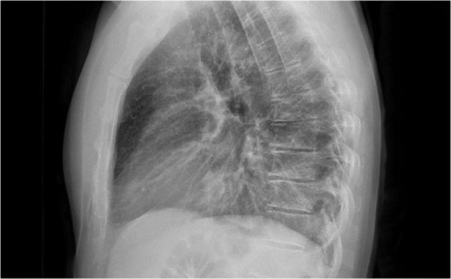
Normal lateral chest X ray

Right hemidiaphragm can be traced back to the posterior wall, but left hemidiaphragm will get silhouetted by the heart ( which lies on the left )
In the previous CXR, the right hemidiaphragm cannot be traced back to the posterior wall, suggesting silhouette sign of posterior part of right hemidiaphragm. This is a right lower lobe pneumonia. Also the lateral CXR should get more radiolucent as it approaches the inferior part, but in the previous picture, it gets more radio-opaque
 |
| Add caption |
Lateral Chest X ray is always a challenge for me, but there are few simple take home points
#1 - The lateral CXR always gets more radiolucent inferiorly; if it gets more radio-opaque, suspect pneumonia in lower lobe ( left/right )
#2 - The retrosternal space is also radiolucent; if it gets radio-opaque, can be retrosternal fat pad, anterior mediastinal masses and RV enlargement
#3 - Remember RALS - right pulmonary artery anterior to right main bronchus and left pulmonary artery
superior to left main bronchus
#4 - Below and posterior to distal trachea ( or origin of bronchus ) is always radiolucent; if it is radio-opaque, suspect pulmonary hypertension ( distension of right/left pulmonary artery ) or lymphadenopathy ( Doughnut sign of Sarcoidosis )
#5 - Right hemidiaphragm can be traced back from anterior to posterior, but left hemidiaphragm can only be traced back from posterior up to the heart; suspect pneumonia in right lower lobe if tracing of right hemidiaphragm is incomplete; suspect pneumomediastinum if the left hemidiaphragm can be traced from front to back ( continuous diaphragm sign )
Reference
Radiology Assistant
Chest X-Ray - Basic Interpretation
http://www.radiologyassistant.nl/en/p497b2a265d96d/chest-x-ray-basic-interpretation.html
What's the diagnosis?
Courtesy of radiology assistant :
Patient has shortness of breath and purulent sputum. Lateral chest X ray is shown here. What does it show?
Sunday, April 6, 2014
Chest radiography !!!
From next week ( and till the end of the month ), my focus will be on the revision of chest radiography, so look forward to it !!!

CAP in kids

Note - please avoid azithromycin if possible !! Doxycycline can cover atypicals very well !!
http://www.medscape.com/viewarticle/820736
Reference
The management of community-acquired pneumonia (CAP) in infants and children older than 3 months of age: clinical practice guidelines by the Pediatric Infectious Diseases Society (PIDS) and the Infectious Diseases Society of America (IDSA). Clin Infect Dis. 2011;53(7):e25–e76
http://pediatrics.aappublications.org/content/128/6/e1677.full
Saturday, April 5, 2014


Menetrier Disease
Learning points from NEJM Case " Unfolding The Diagnosis " April 3 2014
1) Low albumin can be due to 3 reasons

1) Low albumin can be due to 3 reasons
- Liver - look for liver enzymes, bilirubin and coagulation profile abnormalities
- Kidney - look for proteinuria > 3.5 g/day
- GI - malabsorption syndromes ( pancreatitis, celiac disease, crohn's disease and Menetrier disease )
2) Large gastric fold can be due to
- Hyperplasia:- Menetrier disease ( hyperplastic hypoproteinemic gastropathy)
- ZES ( parietal cells hyperplasia due to gastrin stimulation )
- Non hyperplastic mucosal hypertrophy :
- Chronic inflammation - chronic gastritis due to H.pylori
- Infiltration/cancer - sarcoidosis, eosinophilic gastroenteritis, linitis plastica
3) The diagnosis of Menetrier disease can be done by
- CT scan showing thickened gastric folds
- Endoscopy - show thickened folds
- Definitive by biopsy and histological examination
4) Menetrier disease patient commonly presents with GI symptoms and low albumin, if there is viral prodromal symptoms, suspect occult CMV infections
Reference
Unfolding the Diagnosis Gadi Lalazar, M.D., Victoria Doviner, M.D., and Eldad Ben-Chetrit, M.D. N Engl J Med 2014; 370:1344-1348 April 3, 2014

Friday, April 4, 2014


Hepatic encephalopathy
Short post now, what tips the patient with liver failure into hepatic encephalopathy?
ABCDEFGHI
A - Alcohol ( causes more liver destruction, acts as sedative too )
B - Bugs ( infection )
C - Constipation
D - Drugs ( sedatives )
E - Electrolytes ( hypoNa and hypoK )
F - Fluid ( spironolactone esp used to tx ascites )
G - GI bleeding
H - High protein diet ( gets converted to ammonia -> urea cycle )
I - Insufficiency of kidney ( hepatorenal syndrome )
A simpler way is to classify the causes:
Elevated ammonia and other toxic metabolites (ammonia is detoxified by entering the urea cycle in liver ) in blood
- 2 most important causes are GI bleeding and dehydration ( diuretics )
- Others - constipation and high protein diet
- Also remember TIPS ( transjugular intrahepatic portosystemic shunt ) that directly shunts the urea to the SVC bypassing the portal vein
- Hepatorenal syndrome also causes accumulation of toxic metabolites in the blood
Things that can suppress the brain ( neurons activity is already been suppressed by toxins due to ineffective liver metabolism - hypothesis states that GABA is the predominant neurotransmitter involved )
- Sedatives
- Alcohol
Infection is also a common cause ( esp SBP commonly happens in patients with tense ascites )
COPD
And so today finally I have passed one of my hated exams in my medical student life...

COPD ( chronic obstructive pulmonary disorder ) actually refers to two conditions - bronchitis ( the blue bloater ) and emphysema ( the pink puffer ) - of course this is just textbook stuff, real life patient doesn't always present classically


I would think everybody should know about COPD. Smoking is the strongest risk factor for COPD, and global pollution now makes our lungs sicker !!
COPD ( chronic obstructive pulmonary disorder ) actually refers to two conditions - bronchitis ( the blue bloater ) and emphysema ( the pink puffer ) - of course this is just textbook stuff, real life patient doesn't always present classically
Blue bloater - remember bronchitis causes mucus secretion and bronchial inflammation ( which causes it to be edematous and closing the airway ), causing V/Q mismatch and cyanosis ( blue ). And because there is obstruction, CO2 cannot go out ( bloater part )
Pink puffer - emphysema destroys your alveoli together with your capillaries, therefore no V/Q mismatch will happen although ventilation and perfusion both decrease ( pink ). As a result of decreased O2 delivery to capillaries of lung, patient will hyperventilate ( puffer part )
More important than these are:
1) Asthmatic patients can decompensate into COPD
2) COPD is not a local process, it's a systemic inflammatory state
3) Emphysema in young patients should make you consider alpha-1 antitrypsin deficiency - PiZZ homozygous form
Important things to note are:
1) GOLD classification of COPD
- COPD is diagnosed when FEV1/FVC < 70%
- Remember FEV1 is used to stage the disease
- FEV1 > 80% - mild
- FEV1 between 50-80% - moderate
- FEV1 between 30-50% - severe
- FEV1 < 30% or < 50% with resp.failure - very severe
2) Tx options differ according to different stages
- For all stages, SABA is given prn
- For moderate COPD, long acting bronchodilators ( LABA alone or with LAMA - adding together gives addictive effect as they act on different receptors ) are given together with pulmonary rehab
- For severe COPD, inhaled corticosteroid is added
- For very severe COPD, consider O2 therapy and surgical lung reduction
Indications for Long Term O2 Therapy ( LTOT ) are:
1) PaCO2 < 7.3 kPa ( for easy remember < 7 kPa ) / 55mmHg ( if you are reading US sources )
2) If PaCO2 is between 7.3-8 kPa ( 55-59 mmHg ), consider if patient having secondary polycythaemia ( Hct > 45% ) and cor pulmonale
3) What improves Mortality? Remember SOFA
- Smoking cessation ( of course !!! - for me it's the MOST important non-medical treatment )
- Oxygen therapy
- Flu vaccination
- Antibiotics during acute exacerbation
AECOPD ( acute exacerbation of COPD )
Risk factor for severe form AECOPD includes:
1) Previously severe exacerbation : Has previously been admitted to ICU due to severe AECOPD/ near fatal episode
2) use of steroids : patient just starts to use ( indicates at least stage 3 COPD )/ patient never uses it ( decompensation/worsening of COPD )/ patient stops it due to side effects ( COPD is irreversible so once you use it you cannot stop it )
Precipitant for AECOPD is commonly infections ( resp.infections esp )
What to do in AECOPD?
U can remember by this mnemonic AECOPD ( not in order of tx )
A - Antibiotics if suggestive
E - Extra help ( NPPV )
C - Corticosteroids ( oral/IV form has delayed onset of 6 hrs so inhaled form initially may help )
O - Oxygen
P - Pneumococcal and flu vaccination ( done afterwards )
D - Dilators ( SABA prn )
Generally, the order of tx will be
1) Assess ABC - normally B will be the problem , O2 or NPPV
2) IV access and VBG/ABG
3) Bronchodilators for better air entrance and symptomatic relief + oral/IV corticosteroids started ( inhaled corticosteroids can also be given )
4) CXR and sputum C & S after patient has stabilized, antibiotics if needed
There are a few criteria to start antibiotic therapy in AECOPD ( known as Anthonisen Classification )
1) Increased sputum purulence
2) Increased sputum volume
3) Worsening of dyspnea
If 3 are satisfied, abx should be started, if 2 are satisfied, abx recommended if purulence is included, abx is NOT recommended if there is only 1 satisfied
Basically, there are 3 groups of microorganisms causing AECOPD
1) Group A - the usual gram positive bacteria causing CAP and viral causes
- Beta lactams are DOC
2) Group B - the above bacteria but with resistance + gram negative Enterobacteriaceae
- Beta-lactams + beta-lactamase inhibitors are DOC
- Macrolides and resp.fluoroquinolones as alternative
3) Group C - PSA
- Need 2 anti-pseudomonal antibiotics
RFs for severe course of AECOPD includes SEAP
1) Severe COPD
2) Exacerbation is frequent
3) Antibiotics in 3 months ( more risk of abx resistant m/o )
4) Presence of comorbids ( naturally, a CHF patient with AECOPD is more difficult to treat )
These are some of the studies which i think is important to know
- It proves that corticosteroids for 5 days in AECOPD is enough ( REDUCE trial )
- It somehow shows the efficacy of nebulized corticosteroids in AECOPD
- It proves the utility of systemic corticosteroids in AECOPD
Summary
References
Optimizing antibiotic selection in treating COPD exacerbations
Attiya Siddigi and Sanjay Sethi
Int J Chron Obstruct Pulmon Dis. Mar 2008; 3(1): 31–44
http://www.ncbi.nlm.nih.gov/pmc/articles/PMC2528209/
COPD (Chronic Obstructive Pulmonary Disease): What people should Know about COPD http://www.daynightclinic.com/COPD.aspx
Short-term vs conventional glucocorticoid therapy in acute exacerbations of chronic obstructive pulmonary disease: the REDUCE randomized clinical trial.
Leuppi JD1, Schuetz P, Bingisser R, Bodmer M, Briel M, Drescher T, Duerring U, Henzen C, Leibbrandt Y, Maier S, Miedinger D, Müller B, Scherr A, Schindler C, Stoeckli R, Viatte S, von Garnier C, Tamm M, Rutishauser J.
JAMA. 2013 Jun 5;309(21):2223-31
http://www.ncbi.nlm.nih.gov/pubmed/23695200
Steroids in acute exacerbations of chronic obstructive pulmonary disease: are nebulized and systemic forms comparable?
Gunen H1, Mirici A, Meral M, Akgün M.
Curr Opin Pulm Med. 2009 Mar;15(2):133-7
http://www.ncbi.nlm.nih.gov/pubmed/19532028
Optimizing antibiotic selection in treating COPD exacerbations
Thursday, April 3, 2014
Is he wheezing or stridor-ing?

It is always a confusion for me between wheezing vs stridor ( probably mainly because I do not look up enough ). So what is the difference?
From PubMed :

Hmm..so wheezing is stridor? Not quite yet ( subset does not equal to similar )
1) Wheezing is normally heard in expiration ( although can be inspiratory or biphasic ); stridor is the reverse - heard best in inspiration
2) As wheezing is normally heard in expiration, it typically points to diseases in intrathoracic compartment ( i.e bronchi, bronchioles etc ), and stridor typically points to diseases in extrathoracic compartment ( i.e trachea, epiglottis )
3) And because wheezing typically suggests lower airway pathology, it is heard best over in chest, while stridor is heard better in neck ( logically )
4) One more thing is that stridor can be heard without a stethoscope, like this http://www.youtube.com/watch?v=Zkau4yHsLLM and this http://www.youtube.com/watch?v=1Enq2BvX9aw - the characteristic musical sound is typical of stridor
So what is the significance of hearing wheezing and stridor?
Wheezing - think of 3As
- Asthma, Anaphylaxis ( reactive processes )
- Aspiration of foreign bodies
- But remember asthmatic doesn't always wheeze, in fact the disappearance of wheezing in asthmatics equal impending respiratory arrest !! I call it 'paradoxical wheezing' as the airway is become more and more obstructed, the wheeze becomes softer and softer
- CHF can also cause wheezing in kids
- Rare causes like vascular ring which clamps your lower airway ( esp part of trachea located in the thoracic cavity, other symptom includes dysphagia when the esophagus is also clamped down )

Stridor
Epoglottis/Larynx - Acute epiglottitis, croup ( laryngotracheobronchitis ), laryngomalacia, post-intubation laryngeal injury
Trachea - Foreign Body ObstructionReferences
Fundamentals of Lung Auscultation
Abraham Bohadana, M.D., Gabriel Izbicki, M.D., and Steve S. Kraman, M.D. N Engl J Med 2014; 370:744-751
http://www.nejm.org/doi/full/10.1056/NEJMra1302901
Wheezing and stridor Hollingsworth HM. Clin Chest Med. 1987 Jun;8(2):231-40. http://www.ncbi.nlm.nih.gov/pubmed/3304813
Reflection on Lung Adventitious Sounds
First time writing this blog, and I hope this blog will be helpful to those are who are still learning the tips and bits of becoming a doctor ( like me )
Yesterday I was revising about lung sounds. And oh my god i have to say when i came cross these three words ' Crepitation, Rales and Crackles'
How do they differ from each other?
Well i went up for some literature search :

So as you can see the founder of stethoscope was René-Théophile-Hyacinthe Laënnec. He was also the first one to classify adventitious sounds into 5 primary types

As you see, he used the term 'rale' which was translated into 'crepitation' by Forbes - hence the confusion !!!
And if you notice, there are dry vs moist rales - what are those?
Well theoretically speaking, moist rales happens when there are secretions inside your alveoli, and dry rales happen when there is obstruction without secretion. However, at least for me, they sound like the same !! In my opinion, it'll be very good of you to notice that patient has rales and he needs further workup !! Moist vs dry does not matter so much; you will know it's either moist or dry after you know the diagnosis
Recently NEJM has an excellent article discussing about the fundamentals of lung auscultation. According to the author, the term rale has been replaced by crackle
So no more dry vs moist confusion, there is only coarse vs fine crackle ( which in my opinion is appropriate as we can know the level i.e upper vs lower of lung pathology )
Finally, this is my attempted table for the adventitious lung sounds
PS please correct if there's any mistake !!

Hopefully this clears up some of your doubts
References
Fundamentals of Lung Auscultation Abraham Bohadana, M.D., Gabriel Izbicki, M.D., and Steve S. Kraman, M.D. N Engl J Med 2014; 370:744-751
http://www.nejm.org/doi/full/10.1056/NEJMra1302901
Laennec and Auscultation Milli Gupta
http://www.uwomj.com/wp-content/uploads/2013/06/v78n1.61-65.pdf
Classification of Adventitious Sounds The British Medical Journal April 16 1938
http://europepmc.org/articles/PMC2086242/pdf/brmedj04272-0043a.pdf
Subscribe to:
Posts (Atom)